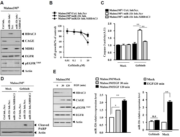
Fig. 7.
miR-326 inhibitor enhances sensitivity to EGFR inhibitors. (A) Malme3MR cells were transfected with the indicated inhibitor (each at 10 nM) along with the indicated siRNA (each at 10 nM). At 48 h after transfection, cell lysates were subjected to Western blot analysis. (B) Malme3MR cells were transfected with the indicated inhibitor (each at 10 nM) along with the indicated siRNA (each at 10 nM). At 24 h after transfection, cells were treated with various concentrations of gefitinib for 24 h, followed by MTT assays. (C) Malme3MR cells were transfected with the indicated inhibitor (each at 10 nM) along with the indicated siRNA (each at 10 nM). At 24 h after transfection, cells were then treated with gefitinib (10 μM) for 24 h, followed by caspase-3 activity assays. **p < 0.005. (D) Malme3MR cells were transfected with the indicated inhibitor (each at 10 nM) along with the indicated siRNA (each at 10 nM). At 24 h after transfection, cells were then treated with gefitinib (10 μM) for 24 h, followed by Western blot analysis. (E) Malme3M cells were treated with EGF (50 ng/ml) for various time intervals. Cell lysates prepared at each time point were subjected to Western blot analysis and qRT-PCR analysis. Malme3M cells were pretreated with gefitinib (10 μM). The next day, cell were then treated with EGF (50 ng/ml) for 2 h, followed by qRT-PCR analysis (right panel). *p < 0.05; **p < 0.005.

