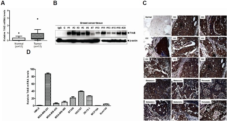
Fig. 1.
Elevated TrkB expression in breast cancer cells and breast cancer patients. (A) The relative expression of TrkB mRNA in 17 human invasive breast carcinoma samples, relative to that of healthy tissues, as determined by quantitative RT-PCR. Expression levels were normalized to 18S mRNA levels. (B) Western blot analysis of TrkB expression in 12 invasive breast carcinoma samples. The β-actin was used as a loading control. (C) Representative immunohistochemical TrkB staining images of normal human breast tissue, infiltrating duct carcinoma, and metastatic carcinoma in the lymph nodes (magnification: 200×). (D) Relative TrkB mRNA expression levels in a panel of eight human nonmetastatic and metastatic cell lines, compared with HMLE cells, as determined by quantitative RT-PCR. The 18S mRNA expression level was used to normalize the TrkB mRNA expression levels.

