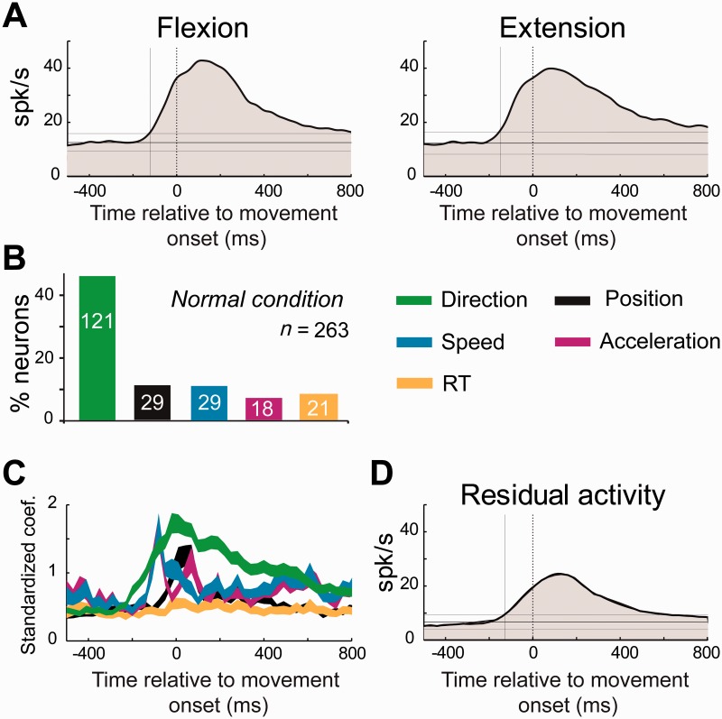
Figure 4.
Time course of kinematic encoding in the neurologically-normal state. (A) Population-averaged spike density functions were aligned on the movement onset (flexion or extension) for all of the movement-related M1 neurons. (B) The fraction of M1 neurons that modulated their activity (permutation test, P < 0.05/54; Equation 1) according to movement direction, hand position, movement speed, acceleration, and reaction time (RT). (C) The population averages of the standardized regression coefficients (β1∼5) at different lags relative to the time of movement onset. The vertical width of each curve represents its standard error of the mean (SEM). (D) The population-averaged residual activity for all movement-related M1 neurons. Residual activity reflects a neuron’s kinematic-independent relationship to movement per se.

