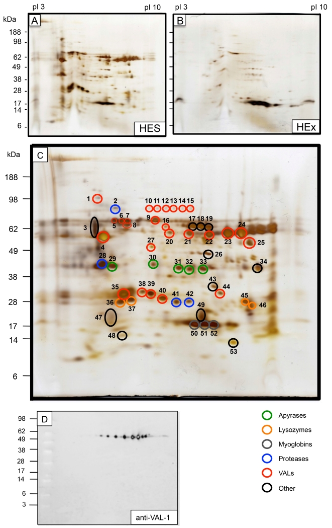
Figure 1. 2-Dimensional analysis of H. polygyrus secreted proteins (HES) and soluble somatic extract (HEx).
A. HES Silver stain
B. HEx Silver Stain. Note that the major spots of 15-18 kDa have previously been identified as myoglobins [24].
C. HES annotated with spots analysed by MS/MS as presented in Table 1
D. Anti-VAL-1 rat polyclonal antibody on Western blot

