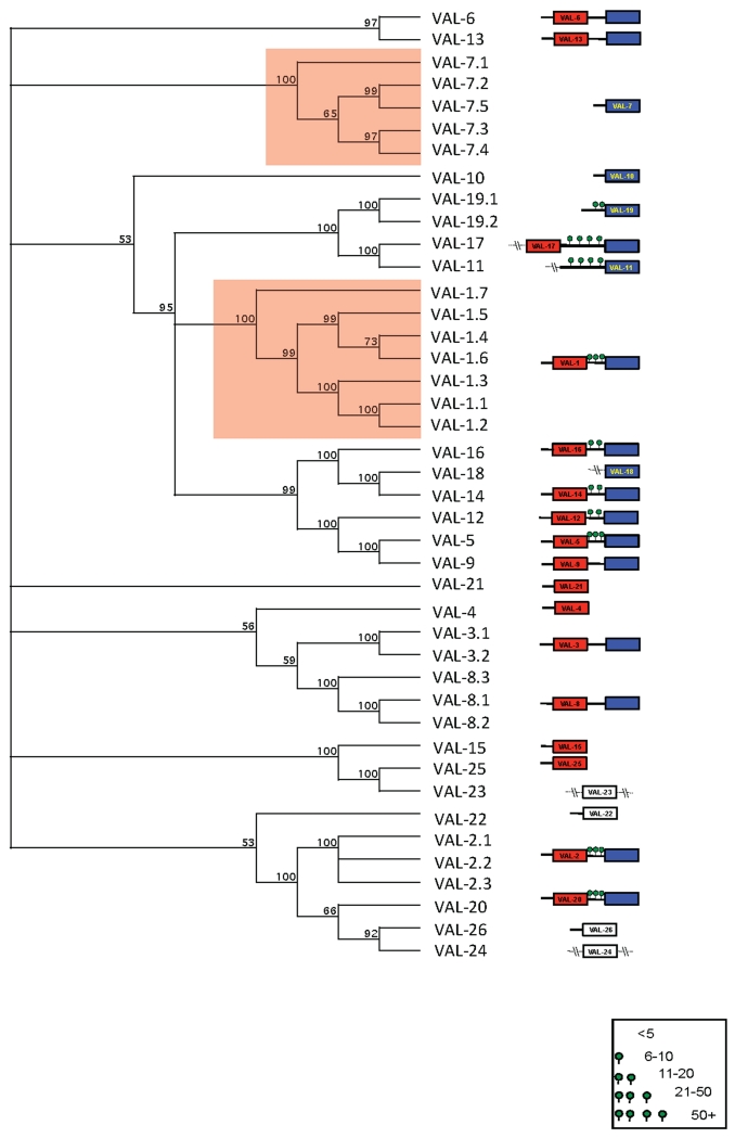
Figure 5. Schematic of relatedness of H. polygyrus VAL-1-25.
The domain structure of Hpb-VAL-1 to -25 are depicted including signal sequences, linker regions (predicted O-glycosylation is indicated with green circles) and SCP homology domains (N-terminal red, C-terminal blue). Single SCP domain proteins are coloured according to whether they are related to N (red) or C-terminal (blue) SCP domains. Divergent sequences equally distinct from both are white. Sequence truncation is indicated by (−\−).

