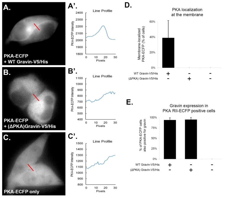
Fig. 3.
PKA RII-ECFP was localized to the cell periphery in AN3 CA cells only when full-length (WT) gravin was present. Representative images and corresponding line-scans (A–C′) show that PKA RII-ECFP localized along the cell periphery only when co-transfected with WT gravin-V5/His. Graph D shows that ~40% of cells expressing PKA RII-ECFP showed ECFP localization at the cell periphery when co-transfected with WT gravin-V5/His. No cells showed PKA RII-ECFP localization at the cell periphery when co-transfected with (ΔPKA) gravin-V5/His, or when expressed in cells containing no gravin. Graph E shows the percentage of PKA RII-ECFP positive cells coexpressing gravin following cotransfection with and without gravin-V5/His constructs. PKA-RII-ECFP positive cells showed a high rate of coexpression with gravin when transfected with gravin-V5/His constructs as indicated by immunofluorescence labeling with a gravin antibody (94% with WT gravin-V5/His; 95% with (ΔPKA) gravin-V5/His). Cells transfected with PKA RII-ECFP only showed no immunofluorescence labeling the gravin antibody. Error bars denote SD.

