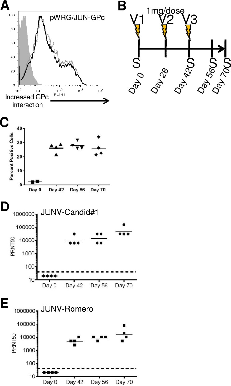
FIG 1.
Generation of anti-JUNV glycoprotein antibodies in rabbits by using i.m. EP. (A) 293T cells were transfected with pWRG/JUN-GPc(opt) and then incubated with anti-JUNV GPc antibody MAb-GB03 (solid black line) or MAb-QC03 (dashed gray line) or the nonspecific control antibody MAb-2G11 (gray shaded area). Cells were subsequently stained with an anti-mouse–Alexa Fluor 488 secondary antibody and analyzed on a flow cytometer. Ten thousand cells were counted per sample, and data were plotted with FlowJo software. (B) Schematic outlining the vaccination of rabbits with pWRG/JUN-GPc(opt). Sera were collected from vaccinated rabbits on days marked with an “S.” Lightning bolts indicate days of vaccination. (C) 293T cells were transfected as described for panel A, except that cells were incubated with rabbit serum collected before (day 0) or after (day 42, 56, or 70) vaccination. The percentages of positive cells were calculated based on values obtained using negative-control serum. (D) JUNV strain Candid#1 was incubated with serially diluted rabbit antisera, and plaque formation was assayed on Vero cell monolayers by neutral red staining. PRNT50 GMTs were calculated based on the plaque formation of virus incubated with the negative-control rabbit antibody. (E) JUNV strain Romero PRNT50 GMTs were determined as described for panel D. Dashed lines indicate the limit of detection for the PRNT assay.

