FIG 6.
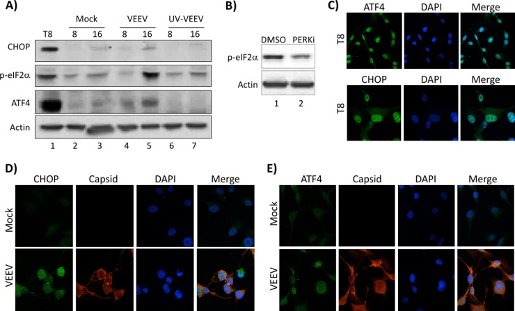
UPR is activated at later time points in VEEV infection. (A) U87MG cells were mock, VEEV, or UV-VEEV infected (MOI, 5), and protein lysates were collected at 4, 8, and 16 hpi (as indicated above each lane). Western blot analysis was performed using antibodies against CHOP, ATF4, p-eIF2α, and actin. T8, U87MG cells treated with tunicamycin for 8 h as a control for UPR induction. Results are representative of those from three independent experiments. (B) U87MG cells were pretreated with DMSO or a PERKi for 2 h prior to infection with VEEV TrD (MOI, 5) and replacement of the medium with drug-containing medium. Western blot analysis was performed using antibodies against p-eIF2α and actin. (C) U87MG cells were treated with tunicamycin for 8 h as a control for UPR induction. (D and E) U87MG cells were mock or VEEV infected (MOI, 5). At 16 h after infection, cells were fixed and probed with DAPI, anti-VEEV capsid, anti-ATF, or anti-CHOP primary antibodies and Alexa Fluor 488- and Alexa Fluor 568-labeled secondary antibodies. Slides were imaged on a Nikon Eclipse TE2000-U fluorescence microscope after immunofluorescence staining. Results are representative of those from two independent experiments.

