FIG 7.
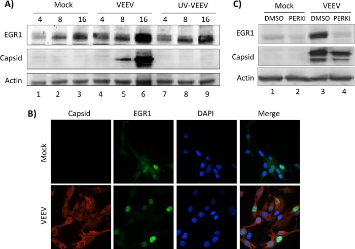
EGR1 is upregulated in infected cells and localizes to the nucleus. (A) U87MG cells were mock, VEEV, or UV-VEEV infected (MOI, 5), and protein lysates were collected at 4, 8, and 16 hpi (as indicated above each lane). Western blot analysis was performed using antibodies against EGR1, capsid, and actin. Results are representative of those from two independent experiments. (B) U87MG cells were mock or VEEV infected (MOI, 5). At 16 h after infection, cells were fixed and probed with DAPI, anti-VEEV capsid, and anti-EGR1 primary antibodies and Alexa Fluor 488- and Alexa Fluor 568-labeled secondary antibodies. Slides were imaged on a Nikon Eclipse TE2000-U fluorescence microscope after immunofluorescence staining. Results are representative of those from two independent experiments. (C) U87MG cells were pretreated with DMSO or a PERKi, infected with VEEV TrD (MOI, 5), and posttreated with drug-containing medium. Protein lysates were collected at 16 hpi. Western blot analysis was performed using antibodies against EGR1, capsid, and actin.

