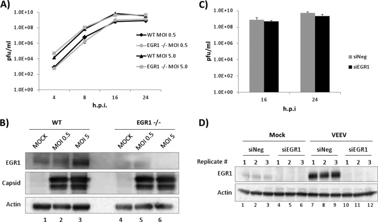
FIG 9.
The loss of EGR1 does not alter VEEV replication kinetics. (A) EGR1−/− and WT MEFs were infected with VEEV (MOI, 0.5 or 5.0). Viral supernatants were collected at 4, 16, and 24 hpi and analyzed by plaque assays. The average from biological triplicates is shown. (B) Protein lysates from EGR1−/− and WT MEFs were separated by SDS-PAGE, and Western blot analysis was performed using antibodies against EGR1, capsid, and actin. (C) U87MG cells were transfected with a negative-control siRNA (siNeg) or siRNA targeting EGR1 (siEGR1). At 48 h posttransfection, cells were infected with VEEV (MOI, 5) or mock infected. Viral supernatants were collected at 16 and 24 hpi, and plaque assays were performed. The average from biological triplicates is shown. (D) Cells were treated as described in the legend to panel C. At 24 h postinfection, cells were collected for Western blot analysis. The results for three independent biological replicates are shown.

