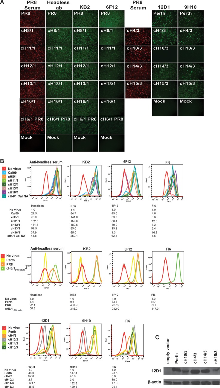
FIG 2.
Conformational stalk epitopes are preserved in cHA of influenza viruses. Stalk-specific antibodies or control serum (PR8 serum) samples were used to probe infected MDCK cells after 16 to 24 h. (A and B) Binding was evaluated using immunofluorescence staining (A) and flow cytometry (B). Immunofluorescence images are representative of results from three samples. Flow cytometry images are representative of results of three experiments for chimeric viruses and two experiments for control viruses Cal09 and Perth. Mean fluorescence intensity (MFI) values for the representative histograms depicted are listed in tables below the respective panels. MFI values were normalized to the “no virus” control value for each panel. ND, not determined. (C) Binding of cHA with group 2 stalks was assessed under denaturing conditions using Western blot analysis performed with antibody 12D1.

