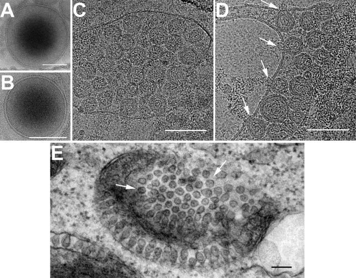
FIG 5.
Electron microscopy of crude mitochondria isolated from mock- and FHV-infected Drosophila cells. (A and B) Cryo-electron microscopy of mitochondria isolated from uninfected (A) and FHV-infected (B) Drosophila cells showing the characteristic double mitochondrial membrane. (C and D) Mitochondria isolated from FHV-infected cells develop spherules after preincubation in RNA replication mix. White arrows in panel D indicate areas where density is seen coming out of the neck region of spherules. (E) Transmission electron microscopy (TEM) of fixed and stained sections from FHV-infected Drosophila cells showing mitochondrial membranes containing spherules perpendicular to the image plane (white arrows). Scale bars = 100 nm.

