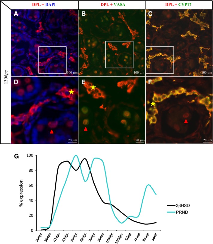
Figure 5.

Expression of Leydig cell markers in goat testis. Dpl (A–F), VASA (B, E) and CYP17 (C, F) immunodetections were performed in a goat testis at 130 dpc. (D–F) photographs correspond to an enlargement of the white rectangles depicted on (A–C) photographs. (G): 3βHSD (black curve) and PRND (blue curve) expression was quantified using real‐time RT‐PCR during testis development in goat and represented on the same graph. At 30 and 36 dpc gonads are not dissected from mesonephros. dpc, days post coïtum; dpp, days post partum; mpp, months post partum.
