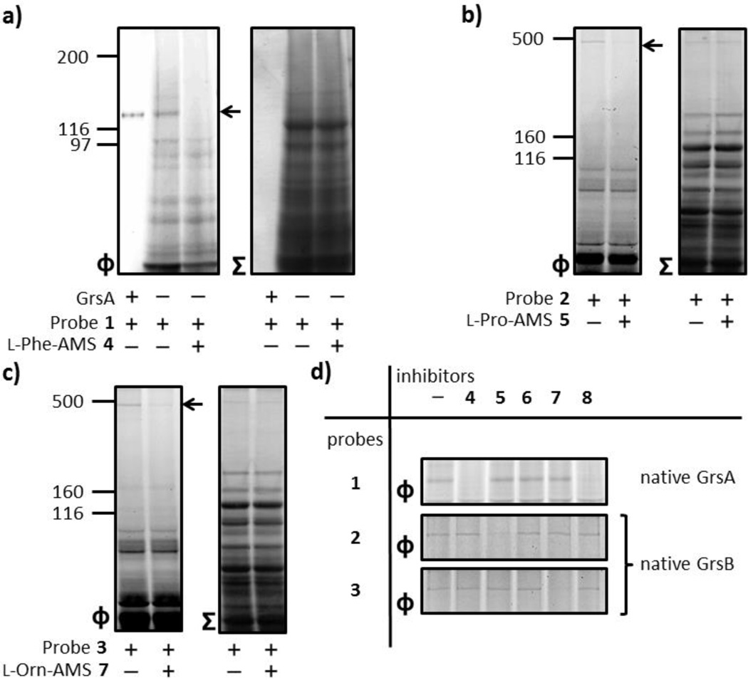
Figure 4.
Proteomic applications of active site-directed proteomic probes 1–3 for A domains. (a) Labeling of endogenous GrsA in an A. migulanus ATCC 9999 cellular lysate by probe 1. The A. migulanus ATCC 9999 lysate (1.5 mg/mL) was treated with 1 µM 1 in either the absence or presence of 100 µM 4. (b) Labeling of endogenous GrsB in the A. migulanus DSM 5759 cellular lysate by probes 2 and 3. The A. migulanus DSM 5759 lysate (1.5 mg/mL) was individually treated with 100 µM 5 and 7 before the addition of individual members of 1 µM probes 2 and 3. (d) Individual labeling of A domains and profiling of A domain functions using a combination of probes 1–3 and inhibitors 4–8. In order to investigate GrsA labeling, the A. migulanus ATCC 9999 lysate (1.5 mg/mL) was preincubated with individual members of inhibitors 4–8 (100 µM) before the addition of 1 µM of probe 1. To evaluate the labeling of GrsB, the A. migulanus DSM 5759 lysate (1.5 mg/mL) was individually treated with 100 µM of inhibitors 4–8 before the addition of individual members of 1 µM probes 2 and 3. For each panel, Φ depicts the fluorescence observed with λex = 532 nm and λem = 580 nm, and Σ displays the total protein content by staining with Coomassie Blue. Full gels (Figure S4) and experimental procedures are provided in the SI.

