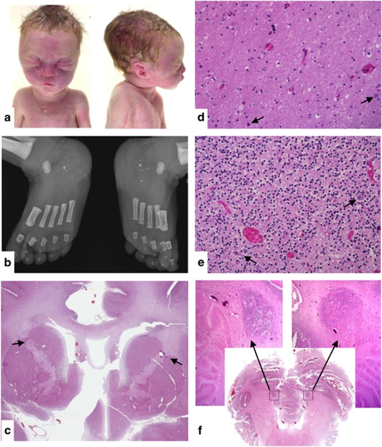
Figure 1.
Fetopathological study of patient no. 1. (a) Facial features: short straight forehead, marked suborbital folds, broad nasal bridge, prominent philtrum, thin upper lip, micrognathia, and abnormally hemmed ears with a small horizontal fold along the upper edge of the helix. (b) Radiographs of the feet: bilateral calcaneal fragmentation and hypermineralisation. (c) Sagittal section through the brain: internal capsule dysmorphism with fusion of anterior caudate nucleus and putamen (black arrows). (d) Cerebral white matter containing ectopic neurons (black arrows). (e) Cerebellar grey matter containing ectopic neurons (black arrows). (f) Sagittal section through the cerebellum showing focal neuronal ectopia.

