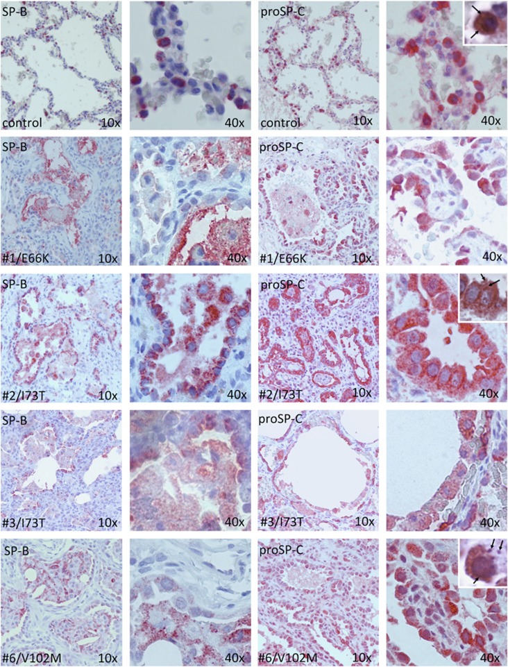
Figure 1.
Lung tissue morphology and surfactant protein immunohistochemistry. Immunohistochemistry staining for SP-B (columns 1 and 2, 10 × and 40 × magnification, respectively) and pro-SP-C (columns 3 and 4) in a 3-month-old control (row 1), three linker domain mutants (row 2–4) and one BRICHOS domain mutant (row 5). Alveolar epithelial type 2 cell hyperplasia is present in all four cases compared with control. In control, proSP-C is expressed in the cytoplasm surrounding clearer-appearing lamellar bodies (row 1, insert, arrows). In cases no. 1–3, proSP-C is diffusely overexpressed in the cytoplasm with a granular pattern (row 3, insert, arrows). In case no. 6, proSP-C is overexpressed with a perinuclear pattern and some scattered aggregates (row 5, insert, arrows). Counterstaining with hematoxylin-eosin.

