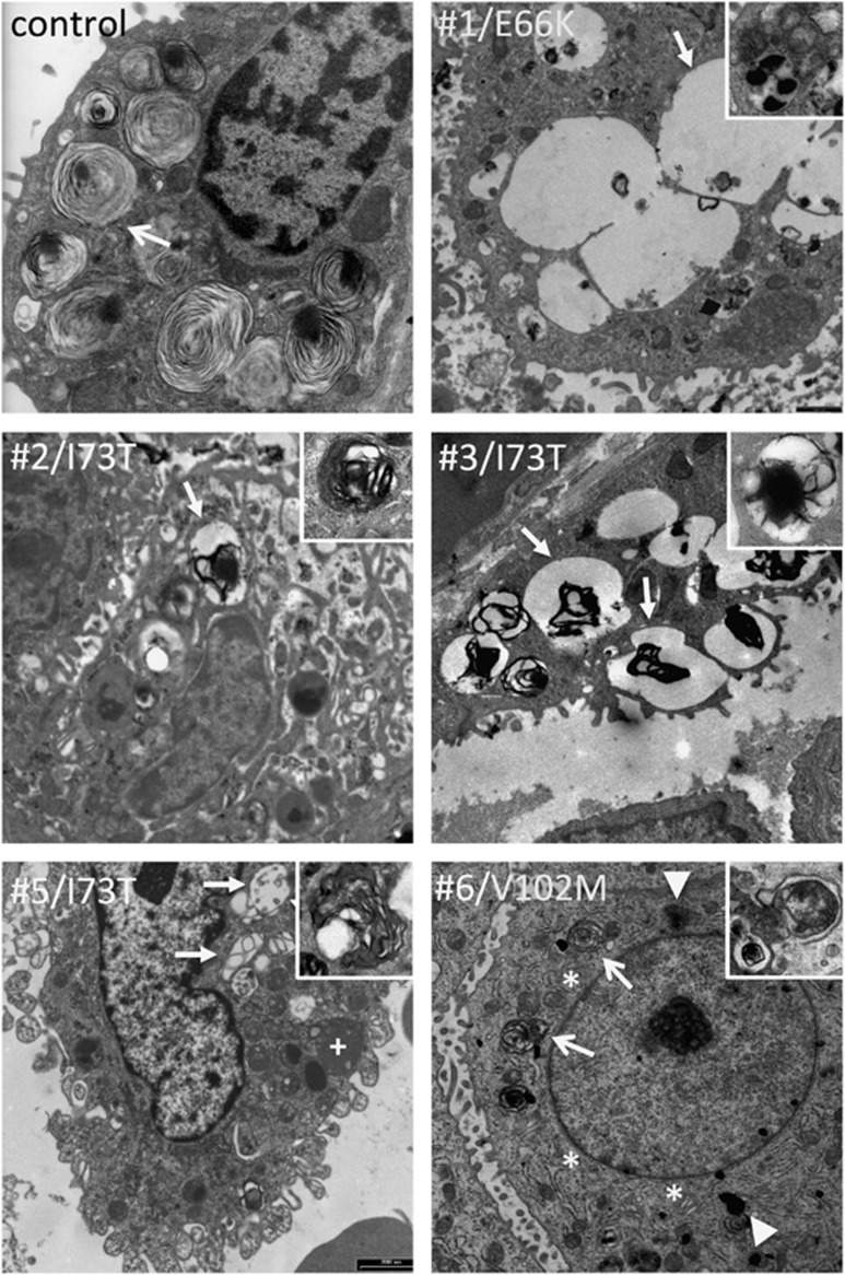
Figure 3.
Type II cell ultrastructure. Type II cell sections in a control subject, four linker domain mutation carriers (cases no. 1–3 and 5) and one BRICHOS mutation carrier (no. 6). Tissue was obtained by autopsy for control and live biopsy for cases no. 1–3 and 6; cells were obtained by bronchoalveolar lavage for case no. 5. In control, numerous mature lamellar bodies with pseudomyelin are present (arrow). Cases no. 1–4 show numerous, large coalescing endosomes with scarce amorphous content (arrow), some lysosomes (+), and few multivesicular bodies (inserts). Case no. 6 shows hypertrophic endoplasmic reticulum (*) compared with control, some cytoplasmic electron-dense deposits (block arrows) and several multivesicular bodies (arrows) with disorganized phospholipid membranes and amorphous content (insert).

