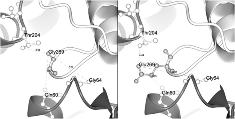
Figure 2.
Structural protein model of wt and mutated ACTG2. Selected part of the ACTG2 model highlighting residue 269 in the actin monomer (light gray) and its relative position to the adjacent actin monomer (deep gray) in the actin polymer. The left panel shows the wt residue Gly269 in close proximity to residues 204, 64 and 60 of the neighboring actin monomer (3.91 Å from Thr204, 7.49 Å from Gly64 and 9.47 Å from Gln60, respectively). The right panel shows the missense variant Glu269 and its side chain that extends further out from the loop. Glutamic acid at position 269 alters distances (and charge) with respect to the closest residues in the neighboring actin monomer (4.44 Å from Thr204, 9.31 Å from Gln64 and 11.05 Å from Gln60, respectively), suggesting an effect on actin monomer interactions.

