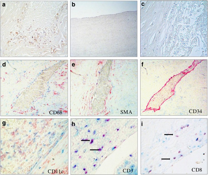
Figure 4.
MCF2L protein is detected in atherosclerotic but not in non-diseased coronary arterial segments and co-localized with CD3 and CD8 Cells. (a) Single staining of MCF2L (brown) in human atherosclerotic tissue. (b) Absence of MCF2L in a normal healthy coronary artery tissue segments. (c) Negative control in atherosclerotic tissue using only the secondary antibody. No background signal is observed. (d) No co-localization of MCF2L (blue) with CD68 (red). (e) No co-localization with smooth muscle alpha-actin (SMA red). (f) No co-localization with CD34 (red). (g) Co-localization of MCF2L (purple) with CD11c antigen-presenting cells, (h) with CD3 type I helper cells and (i) CD8 cytotoxic T cells.

