
Figure 1.
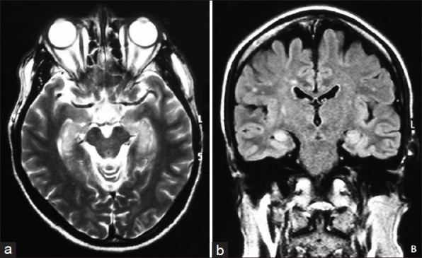
Magnetic resonance imaging of limbic encephalitis associated with anti-GABAB receptor antibodies. (a) T2-weighted magnetic resonance imaging of a patient with γ-aminobutyric acid B receptor abs and limbic encephalitis show increased signal in the mesial temporal lobes; (b) Fluid-attenuated inversion recovery magnetic resonance imaging show increased signal in the mesial temporal lobes.