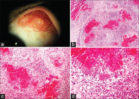
Figure 1.

(a) Slit lamp photograph depicts multiple white-centered cream to yellow colored nodules on the superior bulbar conjunctiva. (b) Scattered eosinophilic materials containing nuclear debris which are surrounded with epithelioid histiocytes, giant cells and lymphocytes demonstrating “zonal granulomatous inflammation”. Eosinophils and plasma cells are diffusely infiltrated in the conjunctival stroma (hematoxylin and eosin, magnification × 100). (c and d) Higher magnification images. Note multinucleated giant cells and scattered infiltration of eosinophils in the conjunctival stroma [hematoxylin and eosin, magnification × 200 (c) and × 400 (d)].
