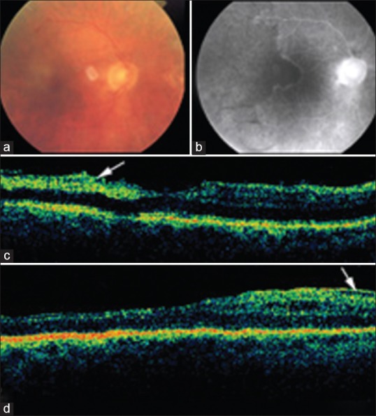
Figure 1.

Case 1: (a) Color fundus photograph of the posterior pole shows juxtafoveal retinal opacification and macular edema. (b) Fluorescein angiography reveals an enlarged foveal avascular zone extending to the temporal retina. (c) Optical coherence tomography (OCT) demonstrated increased reflectivity from the inner retinal layers (arrow). (d) Six months later, OCT demonstrates increased retinal thickness in the temporal fovea with an epiretinal membrane (arrow).
