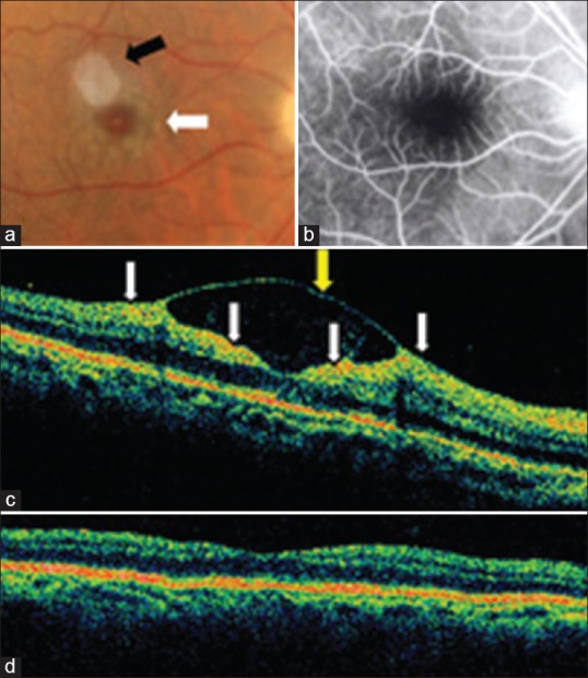
Figure 2.

Case 2. (a) Color fundus photograph shows cotton-wool spots in the fovea and parafoveal area in the right eye (white arrow). The white reflex superotemporal to the fovea is an artifact (black arrow). (b) Fluorescein angiography revealed enlargement of the foveal avascular zone. (c) Optical coherence tomography (OCT) showed internal limiting membrane (ILM) detachment (yellow arrow) in the macular area with increased reflectivity at the inner retinal layer (white arrows) corresponding to retinal ischemia. Note the presence of reflective material within the ILM detachment. This shows that the ILM detachment was not serous and contained inflammatory/proteinaceous fluid (exudative ILM detachment). (d) Nine months later, OCT demonstrates normalization of the foveal contour.
