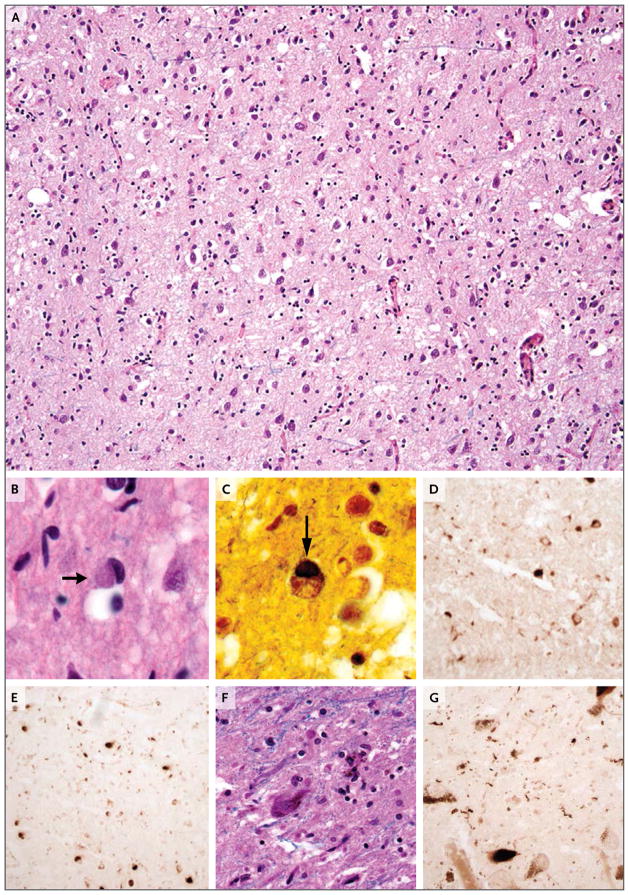
Figure 3. Microscopic Neuropathological Images at Autopsy.
A combination of Luxol fast blue and hematoxylin and eosin staining of the frontal lobe shows attenuation of the neuropil, neuronal loss, and reactive gliosis (Panel A). A Pick body is evident in a cortical neuron (Panel B, arrow). Impregnation with the use of a modified Bielschowsky silver method shows a Pick body (Panel C, arrow). Immunohistochemical staining with phosphospecific antibodies against tau was performed; staining for AT8 (specific for phosphorylation at S199, S202, and T205) (Panel D) and staining for PHF1 (specific for phosphorylation at S396 and S404) (Panel E) show tau-containing inclusions. A combination of Luxol fast blue and hematoxylin and eosin staining shows neuronal loss and extracellular neuromelanin in the substantia nigra pars compacta (Panel F). Immunohistochemical staining for PHF1 shows cytoplasmic inclusions and neuropil threads in the substantia nigra that contain phosphorylated tau (Panel G).

