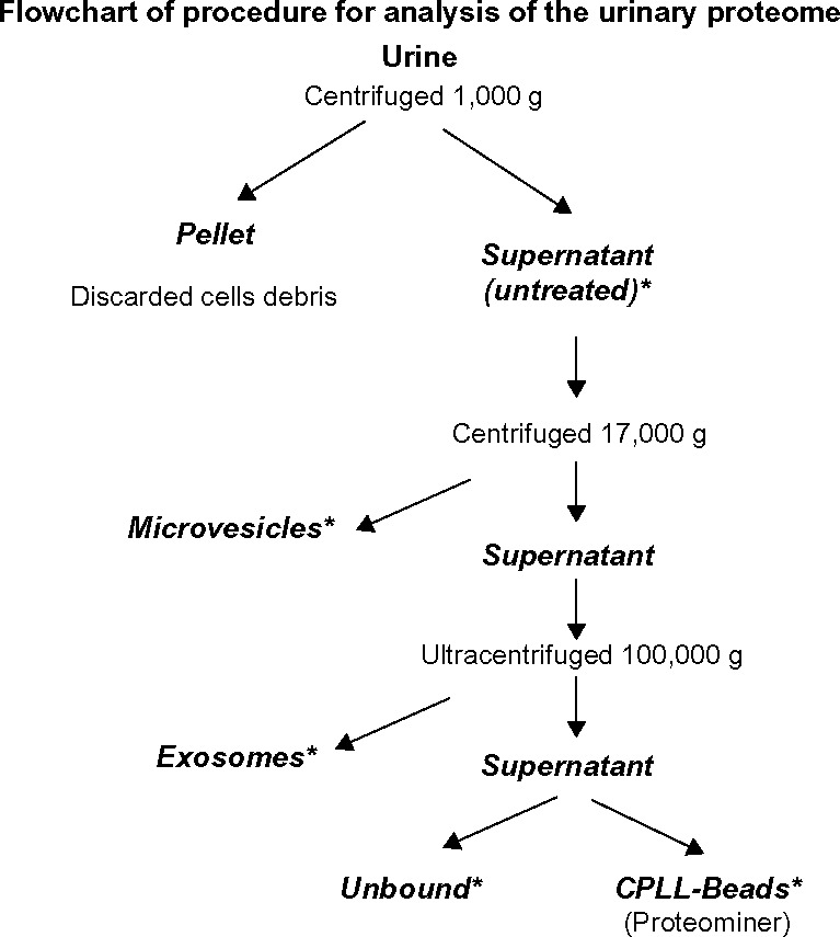
Figure 1.

Proposed flowchart for analysis of the urinary proteome. Urine is centrifuged to separate cell debris. Then, microvesicle and exosome fractions (17,000 and 100,000 × g, respectively) are purified. The supernatant is ultracentrifuged and treated with Proteominer. The five fractions thus obtained (untreated, microvesicles, exosomes, unbound, and CPLL beads) are processed by MS analysis.
Note: *Analysis by Mass spectrometry.
