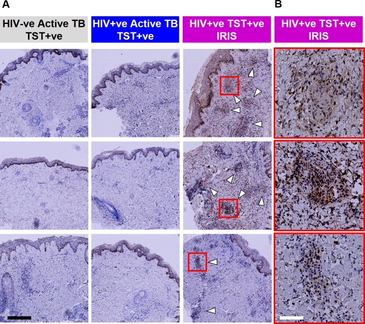
Fig 6. Increased immunostaining for IRF4 in HIV-1 infected patients with unmasking TB-IRIS.
(a) Immunostaining of IRF4 in TST biopsies from three separate patients from each of the study groups indicated (white arrows indicate areas of positive IRF4 staining associated with inflammatory infiltrates). (b) Selected inflammatory cell infiltrates in TB-IRIS cases (indicated by red squares) are shown at higher magnification. Black scale bar = 400 μM and white scale bar = 80 μM.

