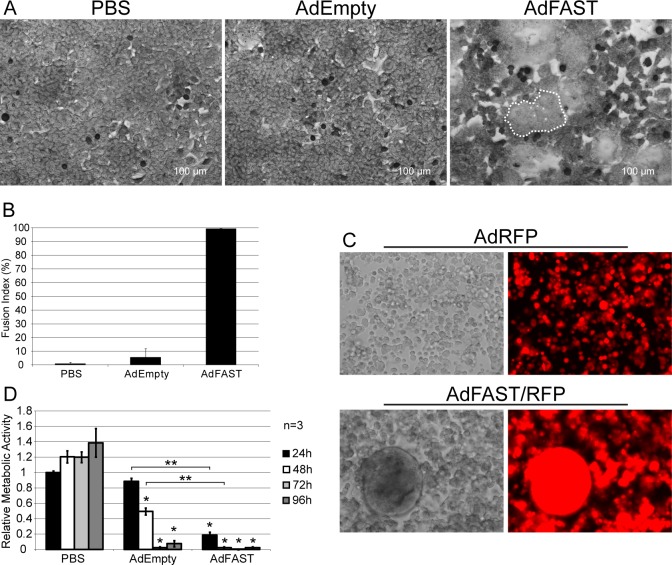
Fig 2. FAST protein expression induces extensive cell fusion in 293 cells.
A) 293 cells were infected with AdEmpty or AdFAST at an MOI of 1 and stained with Giemsa stain 18 h later. Images were captured using bright field microscopy (20x objective). A region of fused cells for the AdFAST-treated cells is outlined with a dotted line, and several nuclei within the syncytium are indicated with asterisks (*) B) Fusion index of 293 cells infected with AdEmpty or AdFAST. The fusion index for two fields of view were determined, and the average with the standard deviation is depicted in the graph. C) 293 cells were infected with AdRFP or AdFAST/RFP at MOI 10 and observed using fluorescence microscopy at 48 h post infection. All images were taken using a 20x objective. D) 293 cells were infected at a MOI of 10 with AdEmpty or AdFAST and relative metabolic activity was determined using an MTS assay over a 96 h interval every 24 h. Experiments were completed in triplicate and the average of three independent experiments is shown (n = 3). Values were normalized to mock infected cells at 24 hpi. Error bars denote the standard error of the mean. *p<0.05 compared to mock infected cells. **p<0.05 when comparing AdFAST to AdEmpty infected cells.

