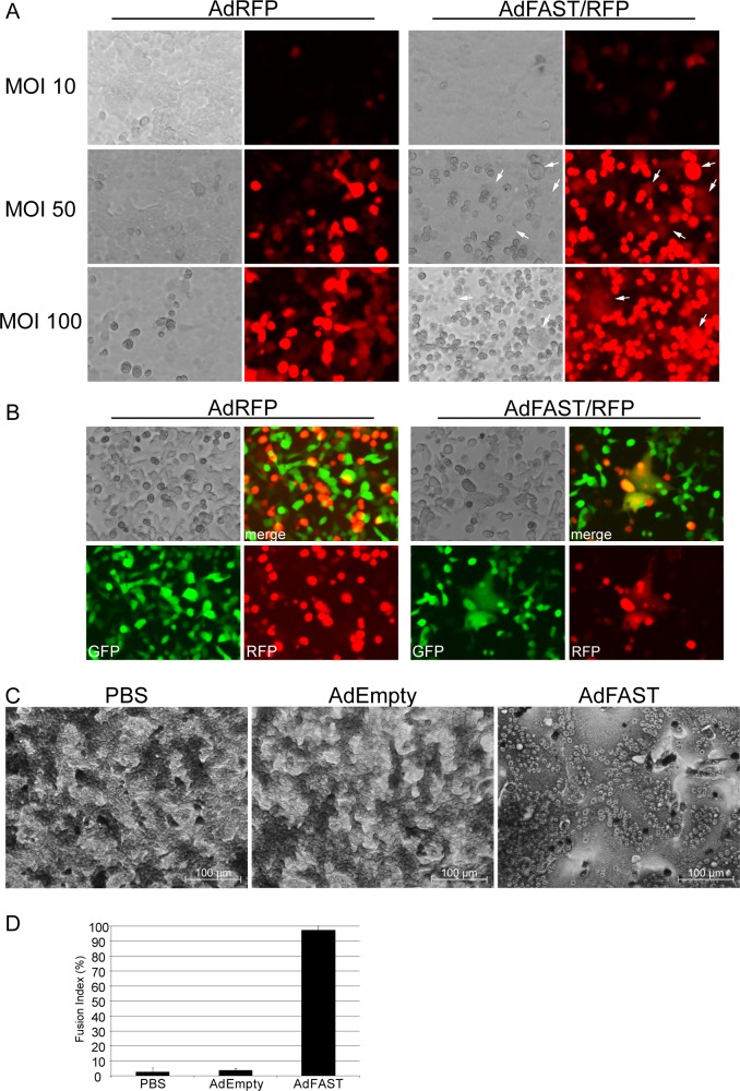
Fig 3. Ad-mediated FAST protein expression induces cell fusion in A549 cells.
A) A549 cells were infected with AdRFP or AdFAST/RFP at an MOI of 10, 50 or 100. Fluorescence microscopy was used to visualize cells 48 hpi (20x objective). Fused cells are indicated by the white arrows. B) A549 cells infected with AdRFP or AdFAST/RFP at an MOI of 10 were cocultured 6 hpi at a 1:1 ratio with A549 cells constitutively expressing GFP. After 72 h of coculturing, cells were visualized using fluorescence microscopy with a 20x objective. The upper right image shows the merged image while the two bottom panels show GFP and RFP fluorescence. C) A549 cells were infected with AdEmpty or AdFAST at an MOI of 50 and stained with Giemsa stain after 48 hpi. Cells were visualized using bright field microscopy with a 20x objective. Two independent experiments were conducted with representative images depicted. D) The fusion index of Giemsa-stained A549 cells infected with AdEmpty or AdFAST at an MOI of 50 was calculated from two fields of view from each independent experiment (n = 2). The average fusion index is shown with the standard deviation.

