
Fig. 7. Confocal microscope images of WT and WT+22 elastic fibers with human retinal pigmented epithelial cells.
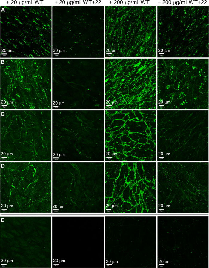
(A to D) Samples were fixed (A) 1 day, (B) 4 days, (C) 7 days, and (D) 10 days after the addition of tropoelastin (20 or 200 μg/ml). (E) Controls (from left to right): sample with no tropoelastin added but stained with anti-elastin mouse antibody and FITC-conjugated anti-mouse antibody; unstained sample with WT (200 μg/ml); sample with WT (200 μg/ml) and stained with FITC-conjugated anti-mouse antibody only; sample with WT (200 μg/ml) and stained with nonspecific mouse IgG and FITC-conjugated anti-mouse antibody. All controls show the absence of visible elastic fibers, indicating the specificity of immunostaining for elastic fibers assembled from exogenous tropoelastin. Scale bar, 20 μm.