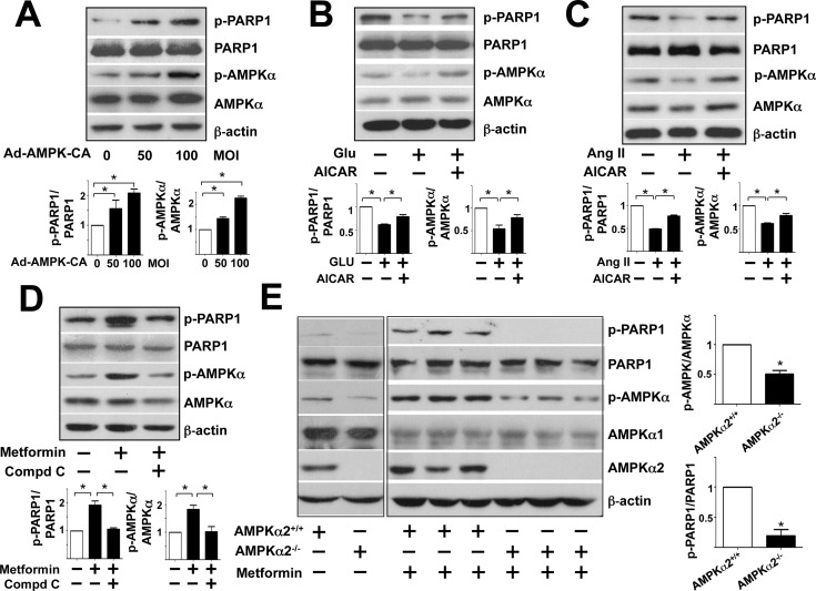
Fig 4. AMPK phosphorylates PARP1 Ser-177 in vitro and in vivo.
Western blot analysis of protein levels in cell lysates and aortic extracts. (A) HUVECs were infected with Ad-AMPK-CA at 50 or 100 multiplicities of infection (MOI) or Ad-null virus at 50 MOI for 24 hr. (B,C) HUVECs were pre-treated with or without AICAR (1 mM) for 30 min before the addition of glucose (30 mM) or Ang II (100 nM) at the indicated concentrations for 4 hr. (D) HUVECs were pre-treated with or without Compound C (15 μM) for 30 min before metformin (5 mM) for 4 hr. (E) AMPKα2+/+ and AMPKα2-/- mice were orally administered with or without metformin (200 mg/kg body weight) and aortas were collected after 12 hr. Data are mean±SD ratio of phospho-PARP1 to total PARP1 and phospho-AMPK to total AMPK from at least 3 experiments in A-D and n = 8 animals in E. *p<0.05 compared with controls.

