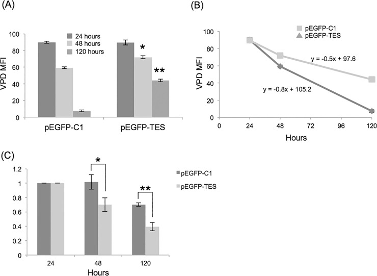
Fig 5. Reduced proliferation of pEGFP-TES transfected NALM6 cells.
(A) Average VPD MFI for GFP-positive, transfected cells at 24, 48 and 120 hours were compared (n = 3). pEGFP-TES transfected NALM6 cells have higher average VPD MFI than GFP-negative (data not shown) or control transfected NALM6 cells, indicating that these cells have undergone fewer cell divisions (unpaired, two-tailed Student’s T-test; * p = 0.003, ** p<0.0001). (B) Proliferation rates of GFP-positive, transfected cells clearly showing reduced proliferation of pEGFP-TES transfected compared to control-transfected cells (line equations were calculated using MS Excel). (C) Reduction in GFP-positive cells over time, calculated relative to 24 hours. Significance was determined between control- and EGFP-TES-transfected cell proportions at 48 (* p = 0.07) and 120 hours (** p = 0.009)(unpaired, two-tailed Student’s T-test).

