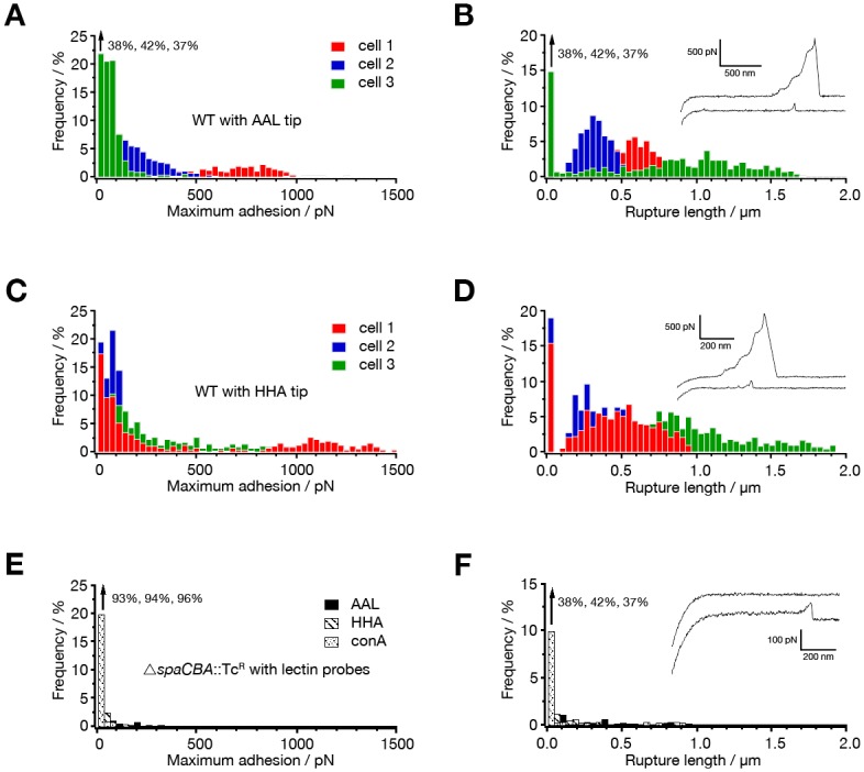
Fig 1. Probing of fucose and mannose residues on L. rhamnosus GG pili using AFM.

Fig 1A and 1C depict the adhesion forces and Fig 1B and 1D the rupture length histograms (n = 1024) obtained in buffer, from the interaction between L. rhamnosus GG wild type and fucose- and mannose-binding lectin probes (AAL and HHA resp.). In Fig 1E and 1F the force data for the interaction of a pili-deficient ΔspaCBA::TcR mutant (CMPG5357) with the two lectin probes are displayed. Insets show representative retraction force curves.
