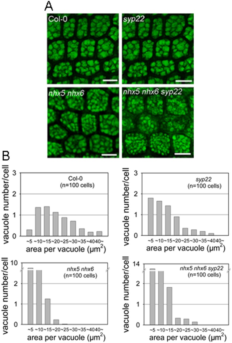
Fig 5. AtNHX5, AtNHX6 and AtSYP22 play an important role in the biogenesis of the PSVs in Aabidopsis.
(A) Morphology of the PSVs. Autofluorescence of the PSVs was visualized with a confocal laser scanning microscope. Bars = 10 μm. (B) Histograms representing a distribution of size and number of vacuoles within a single cell. The X axis indicates the areas of the vacuole. Area of each vacuole was measured from the embryo cells of Col-0 and mutants. 100 cells were measured for PSV analysis.

