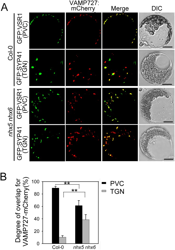
Fig 8. Subcellular localization of AtVAMP727 is altered in nhx5 nhx6.
(A) Subcellular localization of VAMP727:mCherry. The VAMP727:mCherry plasmid was co-transformed with the PVC marker GFP:AtVSR1 and TGN marker GFP:SYP41, respectively, into the leaf protoplasts of Arabidopsis. Fluorescence was visualized by a confocal laser scanning microscope. Bar = 10μm. (B) Quantification of the subcellular localization pattern of AtVAMP727. The overlapping percentage of VAMP727:mCherry signal with GFP:AtVSR1 or GFP:SYP41, respectively, was determined from more than 50 protoplasts obtained from three independent experiments. Error bars represent SD, n = 3. **P≤0.01, by t test.

