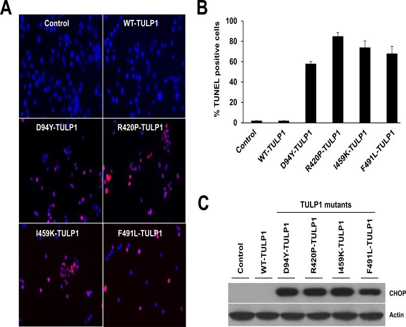
Fig 5. Mutant TULP1 expressing cells undergo apoptosis.
(A) Confocal images of untransfected, WT and mutant TULP1 expressing cells stained with TUNEL (red). (B) Quantification of TUNEL-positive nuclei showed statistically significant differences between untransfected, WT and mutant TULP1 expressing cells (p<0.0001). (C) Western blot analysis using antibodies against the pro-apoptotic transcription factor CHOP protein indicate significant induction of CHOP in mutant TULP1 expressing cell lines as compared to WT-TULP1. Approximately 200 cells from five randomly selected fields of cells from each transfection were evaluated.

