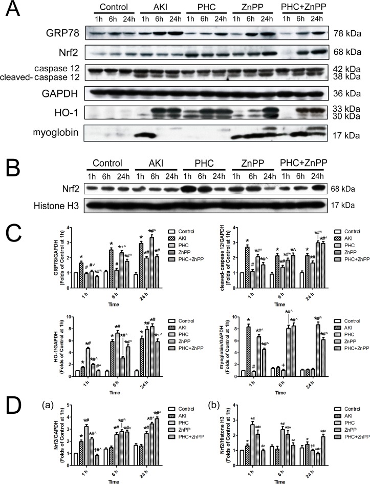
Fig 5. Expression of GRP78, caspase-12, HO-1, myoglobin and Nrf2 in renal tissues by Western blotting.
(A) Expression in total protein of tissues in the boundary area between the renal cortex and medulla, illustrated by original immunoblotting bands and their corresponding molecular weights. The housekeeping protein GAPDH served as reference. (B) Expression of Nrf2 in nuclear protein of tissues in the boundary area between the renal cortex and medulla. Histone H3 served as reference. Each experiment was repeated at least three times. (C) Densitometrical quantitative assessment of the expression of GRP78, caspase-12, HO-1 and myoglobin in total protein. (D) Densitometrical quantitative assessment of the expression of Nrf2 a) in total protein, b) in nuclear protein. Each bar represents mean ± SD. Compared with simultaneous group: * P<0.01 or † P<0.05 vs. Control; # P<0.01 or + P<0.05 vs. AKI; ^ P<0.01 or ˅ P<0.05 vs. PHC.

