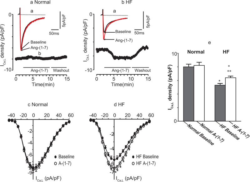
Figure 2.
Effects of Ang-(1-7) on ICa,L in normal and ISO-induced HF myocytes. (a) Examples of original current traces were elicited by depolarization pulses from a holding potential of −80 mV to 0 mV for 200 ms (with a brief prepulse to −45 mV, 60 ms). Ventricular myocytes were initially superfused with external solution. After a stable steady peak ICa,L was recorded, Ang-(1-7) 10−5 M was superfused. The superimposed current tracings were recorded before and after exposure to Ang-(1-7). (b) The time course of peak ICa,L measured every 10 s and plotted as a function of time (in min). Horizontal bars below the graph indicate the time and duration of the application of Ang-(1-7). The voltage protocol was the same as in (a). (c) Current–voltage (I-V) relationship of ICa,L in the absence and presence of Ang-(1-7) in 8 normal myocytes. The cells were depolarized from a holding potential of −80 mV to a test potential from −40 to +60 mV in 5 mV step increments. (d) I-V relationship for 8 HF cells, measured under the same experimental conditions as in (c) * indicates that the difference between before and after drug is statistically significant (p < 0.05). (e). Group means of ICa,L responses to Ang-(1-7) in normal and HF myocytes (n = 10 and 26, respectively). Values are mean (± SEM); *p < 0.01 versus baseline; **p < 0.05, Ang-(1-7) versus baseline. The results showed that, in normal myocytes, Ang-(1-7) has no significant effect on ICa,L. In contrast, in HF myocytes, Ang-(1-7) caused significant increases in ICa,L.
HF, heart failure; ICa,L, L-type calcium current; SEM, standard error of the mean.

