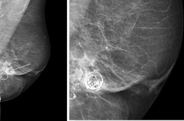
Fig.1.

A, Left MLO and magnified views. Mixed fibroglandular parenchyma, scattered calcifications (arrows and circle) with a wide variety of appearances can be seen in the left breast. US scan showed cystic lesions with heterogenous appearance typical of fat necrosis reported as M2.
