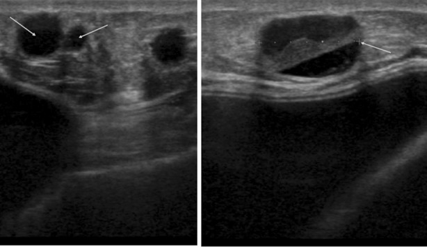
Fig.3.

Clinically palpable lump above WLE scar 4 years after grafting (140 ml) in 2008. Mammograms reported as R2. Ultrasound scan showed 14 mm mixed echogenicity lesion reported as likely to be fat necrosis/oil cyst U2, but in view of the solid component ultrasound guided core biopsy performed which confirmed fat necrosis.
