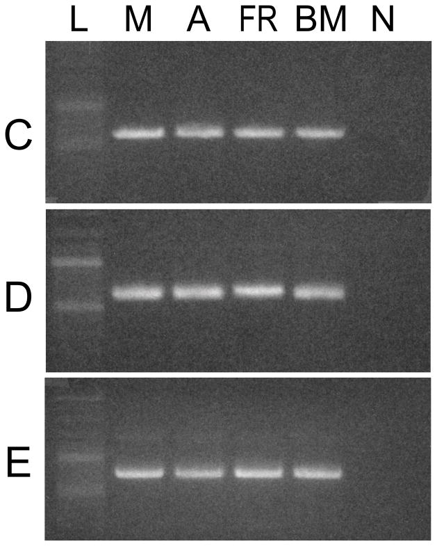
Fig. 5.
Expression of isoforms C, D, and E in muscular tissues of Octopus bimaculoides. PCR was used with specific isoform C, D, and E reverse primers to detect isoforms in O. bimaculoides tissues. The top panel shows the distribution of isoform C; the middle panel that of isoform D; and the bottom panel that of isoform E. L, ladder; M, mantle; A, arm; FR, funnel retractor; BM, buccal mass; N, non-template control.

