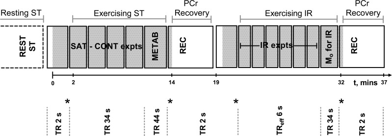
Fig. 1.
Schematic representation of the 31P magnetic resonance spectroscopy (MRS) exercise protocol. Solid lines symbolize sequence blocks, gray shaded regions correspond to when exercise occurred. Time from onset of exercise is illustrated by the timeline. In the first exercise section, once exercising steady-state conditions were met, spectra were obtained with saturation of the γ-ATP resonance and then control saturation placed equidistant to the inorganic phosphate (Pi) resonance (SAT-CONT expts). Spectra were also obtained with a long repetition time (TR) of 44 s for calculation of metabolite concentrations (METAB). Following exercise cessation a phosphocreatine (PCr) recovery measurement (REC) was used to assess the immediate end-of-exercise oxidative ATP synthesis rate. Within the second exercise, once in steady state, the inversion recovery data were acquired with varying times between the inversion and subsequent excitation pulse (TIs) (IR expts) with an effective TR of 6 s, and a measure of Mo was also obtained. The four stars represent comparison sites for steady-state conditions.

