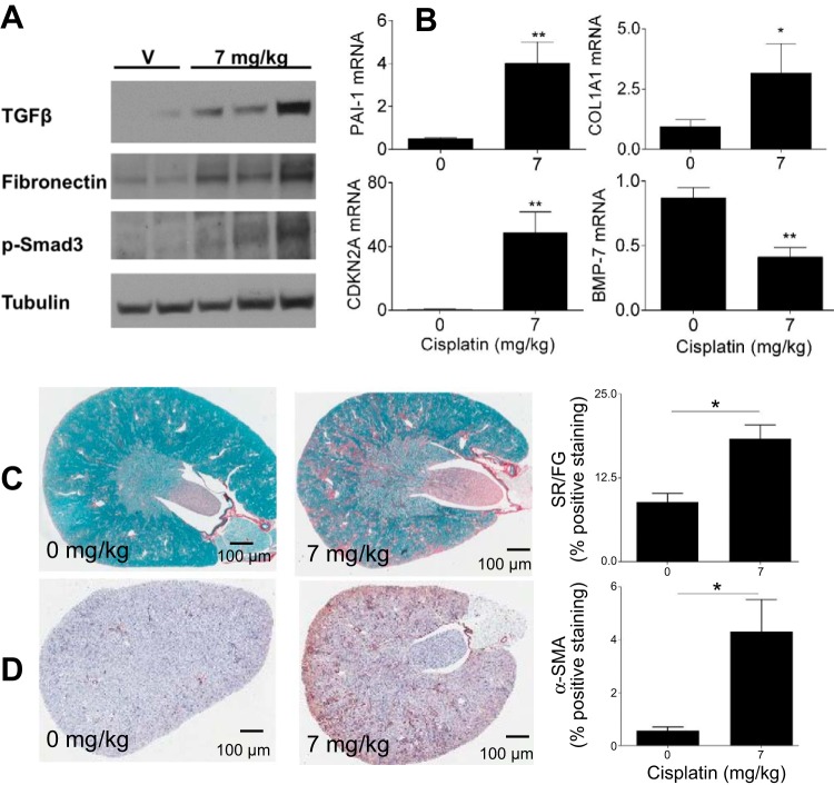
Fig. 6.
Assessment of fibrosis and fibrotic markers. Eight-week-old male FVB mice were injected (intraperitoneally) with saline vehicle or cisplatin (7 mg/kg) once a week for 4 wk (repeated dosing model). Animals were euthanized 72 h after the last injection. A: markers of fibrosis in the kidney cortex assessed via Western blot analysis. TGF-β, transforming growth factor-β; p-Smad, phosphorylated Smad. B: measurement of mRNA levels of fibrotic markers in the kidney cortex as assessed via quantitative RT-PCR. PAI-1, plasminogen activator inhibitor-1; COL1A1, collagen type I-α1; CDKN2A, cyclin-dependent kinase inhibitor 2A (CDK)N2A; BMP-7, bone morphogenetic protein-7. C and D: Sirius red and fast green (SR/FG) staining of kidney sections and quantitation of staining (C) as well as α-SMA immunohistochemical staining in the kidney cortex and quantitation of staining (D). Data are expressed as means ± SE; n = 5–10. Statistical significance was determined by Student's t-test. *P < 0.05; **P < 0.01.

