Fig. 1.
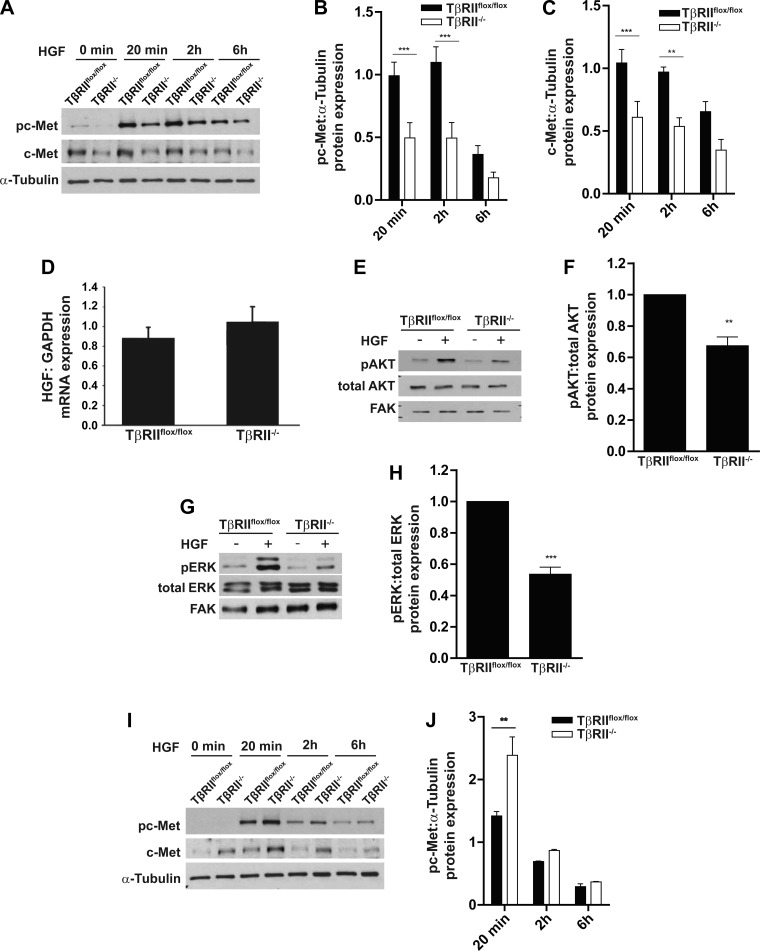
Transforming growth factor-β (TGF-β) receptor type II (TβRII)-deficient (TβRII−/−) proximal tubule (PT) cells have impaired hepatocyte growth factor (HGF) signaling. A–C: TβRIIflox/flox (floxed control) and TβRII−/− PT cells were treated with HGF (40 ng/ml) for 20 min, 2 h, and 6 h and immunoblotted for c-Met expression and c-Met phosphorylation (pc-Met), which were quantified using α-tubulin as a loading control. D: quantitative PCR (qPCR) analysis of HGF transcription by TβRIIflox/flox and TβRII−/− PT cells normalized to GAPDH. E–H: TβRIIflox/flox and TβRII−/− PT cells were treated with HGF (40 ng/ml) for 20 min and immunoblotted for AKT and ERK phosphorylation (pAKT and pERK), which was quantified. FAK, focal adhesion kinase. I and J: lysates of TβRIIflox/flox and TβRII−/− cortical fibroblasts were treated with HGF (40 ng/ml) for 20 min, 2 h, and 6 h and immunoblotted for c-Met phosphorylation, which was quantified using α-tubulin as a loading control. Values are means ± SE of results from 3 separate experiments. **P < 0.01; ***P < 0.0001.

