Fig. 1.
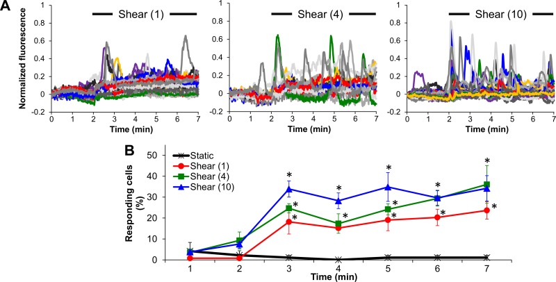
Shear stress causes endothelial cell (EC) intracellular free calcium concentration ([Ca2+]i) transient increases compared with static. A: characteristic normalized fluo-4 fluorescence signals vs. time during a 2-min static followed by a 5-min shear period at either 1, 4, or 10 dyn/cm2 (each colored line corresponds to a single cell in a microscope field of view; 10 colors are being repeated). B: responding cells (%) plotted every minute (at the end of each minute) during either a 7-min static period or a 2-min static period followed by a 5-min shear period (1, 4, or 10 dyn/cm2). Data are means ± SE (n = 4–8 independent experiments). *P < 0.05 vs. its corresponding (preshear) static period (average of the first 2-min data points).

