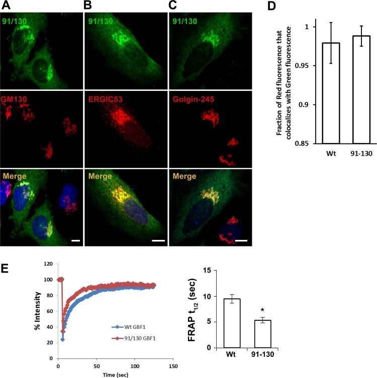
Fig. 2.
91/130 targets to the Golgi and rapidly exchanges between membranes and cytosol. A–C: HeLa cells were transfected with GFP-91/130, and after 24 h cells were processed for immunofluorescence (IF) to localize the GFP and the indicated markers. GFP-91/130 colocalizes extensively with GM130 and ERGIC53 but less so with Golgin-245, indicating localization with a bias toward the early Golgi compartments. Bars, 10 μm. D and E: HeLa cells were transfected with GFP-GBF1 or GFP-91/130, and after 24 h cells were processed for IF to localize the GFP and GM130 (D) or regions within the Golgi were subject to fluorescence recovery after photobleaching (FRAP; E). Mander's overlap coefficient (M1) for the red channel was quantified. The M1 for GFP-GBF1 against GM130 was 0.979 ± 0.026 (mean ± SD), while the M1 for the 91/130 mutant was 0.988 ± 0.013. These values are not statistically different (t-test, P = 0.0646). E: HeLa cells were transfected with GFP-GBF1 or GFP-91/130, and after 24 h regions within the Golgi were subject to FRAP. Averages of 17 GFP-GBF1 and 18 GFP-91/130 determinations are plotted on left. The half-life (t1/2) values were obtained from the exponential aspect of each graph, and averages are plotted on right. GFP-GBF1 FRAP t1/2 was 9.4 ± 0.8 s (average ± SE), and 91/130 FRAP t1/2 was 5.3 ± 0.6 s. There was a statistically significant difference between the t1/2 values as measured by a t-test (*P = 0.0003).

