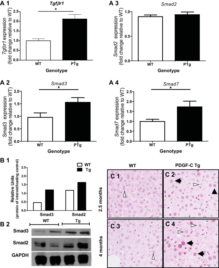
Fig. 1.
Evidence of transforming growth factor-β (TGFβ) signaling in livers of PDGF-C transgenic (PTg) mice. Analysis of gene expression (A), protein (B), and phosphorylated Smad2/3 (C) was performed using livers from PTg mice compared with wild-type (WT) mice (8–9 wk of age, n = 6–7). A: TGFβ1 receptor (Tgfβr1) (A1), Smad3 (A2), and Smad7 (A4) gene expression is higher in PTg mice, while Smad2 (A3) expression did not change. Total RNA was prepared from livers of PTg or WT littermates. Real-time PCR was performed to determine mRNA levels of Tgfβr1, Smad2, Smad3, and Smad7 as described in materials and methods. Data are represented as fold change compared with a WT animal. B: immunoblot analysis of Smad2 and Smad3 indicates that Smad3 is elevated in PTg mice liver tissue, while Smad2 levels were similar to WT mice. The bar graph represents the average of densitometry value for two animals after normalization to GAPDH levels. C: phosphorylated Smad2/3 is present in hepatocyte nuclei, and nonparenchymal cells (NPCs) in livers from PTg (C2 and C4), but not WT (C1 and C3) mice. Immunohistochemical analysis was performed on 2-mo-old (C1 and C2), and 4.5-mo-old (C3 and C4) mice as described in materials and methods. Both stained (black arrowheads) and unstained (white arrowheads) hepatocyte nuclei can be seen in the same field. *P < 0.05.

