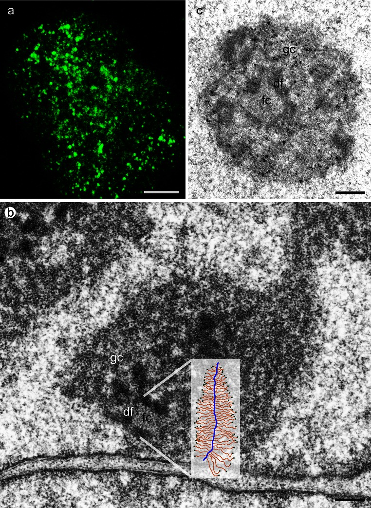
Fig. 1.
a BrU incorporation to visualize nascent transcripts, HeLa cell, confocal image (projection) Bar 5 µm b Nucleolus of HeLa cell, sketch of Christmas tree in relation to the fibrillar complex where transcription takes place (inset), TEM Bar 1 µm c In situ hybridization to detect rRNA which is present in df and gc, HeLa cell, TEM, Bar 1 µm, fc fibrillar center, df dense fibrillar component, gc granular component

