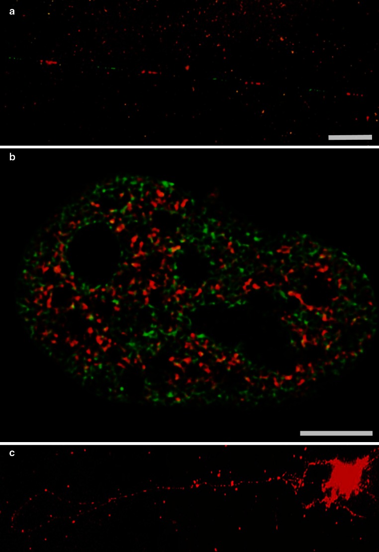
Fig. 2.
a Detection of a fragment of the transcription unit (red) and of the intergenic spacer (green) of rDNA, FISH on stretched DNA fibers, nuclear halo preparation, Bar 5 µm, b HeLa cell expressing histones H2B (green) and histone H3 variant H3.3 (red) which has been associated with transcriptional activity (Ahmad and Henikoff 2002), note that nucleoli are largely devoid of signal, structured illumination imaging, Bar 5 µm, c FISH with a probe covering the entire rDNA repeat showing an extracted rDNA loop, nuclear halo preparation

