
Fig. 1.
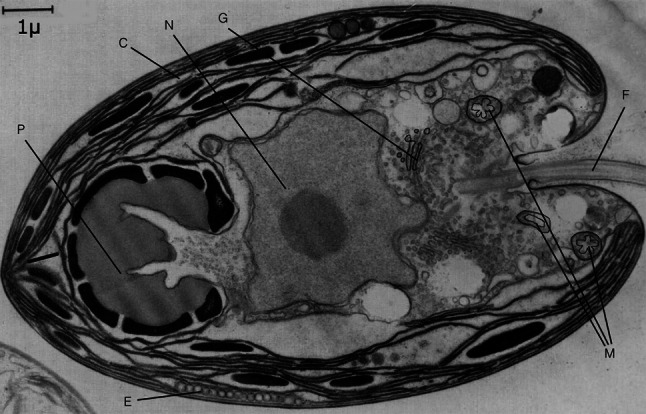
A section of a Tetraselmis cell showing the disposition of the nucleus (N), Golgi bodies (G), mitochondria (M), cup-shaped chloroplast (C), pyrenoid (P), eyespot (E) and flagella (F). Redrawn from (Manton and Parke 1965)
Official websites use .gov
A
.gov website belongs to an official
government organization in the United States.
Secure .gov websites use HTTPS
A lock (
) or https:// means you've safely
connected to the .gov website. Share sensitive
information only on official, secure websites.
A section of a Tetraselmis cell showing the disposition of the nucleus (N), Golgi bodies (G), mitochondria (M), cup-shaped chloroplast (C), pyrenoid (P), eyespot (E) and flagella (F). Redrawn from (Manton and Parke 1965)