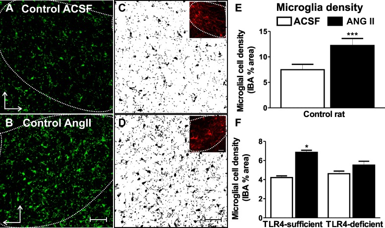
Fig. 1.
ANG II induces microglial activation within the paraventricular nucleus (PVN) of the hypothalamus of control rats and mice, an effect that is blunted in the absence of functional Toll-like receptor (TLR)4. A and B: confocal images of microglial marker ionized calcium-binding adaptor molecule (IBA)1 staining in the presence of artificial cerebrospinal fluid (aCSF; A) and ANG II (1 μM, 60 min; B) in control rats. C and D: IBA1 signal-subtracted threshold used for quantification. Insets show vasopressin stain used as an anatomic marker to trace the region of interest (PVN). E and F: summary data showing the ANG II-induced increase in microglial cell density in control rats (n = 10; E) and TLR4-sufficient mice (n = 5; F). Microglial cell density was not altered in TLR4-deficient mice in the presence of ANG II (n = 3; F). ***P < 0.0001 and *P < 0.05 vs. aCSF. The vertical arrow points dorsally and the horizontal arrow points medially. Scale bars = 50 μm.

